Fysiologibogen 2. udgave
Her finder du figurer fra Fysiologibogen 2. udgave. Du kan læse mere om bogen her.
© Kopiering fra denne hjemmeside må kun finde sted på institutioner eller virksomheder der har indgået aftale med Copydan Tekst & Node og kun inden for de rammer der er nævnt i aftalen.

Figur 2
Na+/K+-ATPasens placering i membranen.
Illustration: Henning Dalhoff
ISBN 978-87-90363-84-0 · © Nucleus Forlag ApS.

Figur 3
Cellen med vigtige organeller.
Illustration: Henning Dalhoff
ISBN 978-87-90363-84-0 · © Nucleus Forlag ApS.

Figur 4
Et phospholipidmolekyle.
Illustration: Henning Dalhoff og Elin Steffensen, Griffle
ISBN 978-87-90363-84-0 · © Nucleus Forlag ApS.

Figur 5
Cellemembranens phospholipiders bevægelse med cholesterol.
Illustration: Henning Dalhoff og Birthe Møller Nielsen
ISBN 978-87-90363-84-0 · © Nucleus Forlag ApS.

Figur 6
Proteiner i cellemembranen.
Illustration: Henning Dalhoff
ISBN 978-87-90363-84-0 · © Nucleus Forlag ApS.

Figur 7
Osmose.
Illustration: Henning Dalhoff
ISBN 978-87-90363-84-0 · © Nucleus Forlag ApS.

Figur 8
Røde blodceller i tre forskellige osmolariteter.
Illustration: Henning Dalhoff
ISBN 978-87-90363-84-0 · © Nucleus Forlag ApS.

Figur 9
Model af den spændingsstyrede Na+-kanal.
Illustration: Henning Dalhoff
ISBN 978-87-90363-84-0 · © Nucleus Forlag ApS.

Figur 10
Model af receptorstyrede ionkanaler.
Illustration: Henning Dalhoff
ISBN 978-87-90363-84-0 · © Nucleus Forlag ApS.

Figur 11
Agres eksperiment med celler som henholdsvis indeholdt og manglede aquaporiner.
Illustration: Henning Dalhoff
ISBN 978-87-90363-84-0 · © Nucleus Forlag ApS.

Figur 12
a. Todimensionel model af aquaporin 1.
Illustration: Henning Dalhoff
ISBN 978-87-90363-84-0 · © Nucleus Forlag ApS.

Figur 13
Vandets passage gennem aquaporinen AQP 1.
Illustration: Henning Dalhoff
ISBN 978-87-90363-84-0 · © Nucleus Forlag ApS.

Figur 14
Faciliteret diffusion ved hjælp af specielle transportproteiner.
Illustration: Henning Dalhoff
ISBN 978-87-90363-84-0 · © Nucleus Forlag ApS.

Figur 15
Forskellige former for aktiv transport.
a. Uniporter.
b. Symport cotransporter.
c. Antiport cotransporter.
Illustration: Henning Dalhoff
ISBN 978-87-90363-84-0 · © Nucleus Forlag ApS.

Figur 16
Sekundær aktiv transport.
Illustration: Henning Dalhoff
ISBN 978-87-90363-84-0 · © Nucleus Forlag ApS.

Figur 17
Na+/K+-pumpens otte trin.
Illustration: Henning Dalhoff
ISBN 978-87-90363-84-0 · © Nucleus Forlag ApS.

Figur 18
Endocytose og exocytose.
Illustration: Henning Dalhoff
ISBN 978-87-90363-84-0 · © Nucleus Forlag ApS.

Figur 19
Model af insulinreceptoren.
Illustration: Henning Dalhoff
ISBN 978-87-90363-84-0 · © Nucleus Forlag ApS.

Figur 20
Indirekte respons med den G-proteinkoblede receptor.
Illustration: Henning Dalhoff
ISBN 978-87-90363-84-0 · © Nucleus Forlag ApS.

Figur 21
Model af den G-proteinkoblede receptor.
Illustration: Henning Dalhoff
ISBN 978-87-90363-84-0 · © Nucleus Forlag ApS.

Figur 22
Intracellulær receptor.
Illustration: Henning Dalhoff
ISBN 978-87-90363-84-0 · © Nucleus Forlag ApS.

Figur 23
a. Nikotin som agonist.
b. Nalone som agonist.
Illustration: Henning Dalhoff
ISBN 978-87-90363-84-0 · © Nucleus Forlag ApS.

Figur 24
Gap junctions mellem celler.
Illustration: Henning Dalhoff
ISBN 978-87-90363-84-0 · © Nucleus Forlag ApS.

Figur 25
a. Tværsnit af hjernen.
b. Hjernen.
Illustration: Henning Dalhoff
ISBN 978-87-90363-84-0 · © Nucleus Forlag ApS.

Figur 26
Oversigt over centralnervesystemet og det perifere nervesystem.
Illustration: Henning Dalhoff
ISBN 978-87-90363-84-0 · © Nucleus Forlag ApS.

Figur 27
Det autonome nervesystem består af sympatikus og parasympatikus.
Illustration: Henning Dalhoff
ISBN 978-87-90363-84-0 · © Nucleus Forlag ApS.

Figur 28
a. Et typisk neuron.
b. Schwannsk celle.
Illustration: Henning Dalhoff
ISBN 978-87-90363-84-0 · © Nucleus Forlag ApS.

Figur 29
Eksempler på forskellige typer af neuroner.
Illustration: Henning Dalhoff
ISBN 978-87-90363-84-0 · © Nucleus Forlag ApS.

Figur 30
En nerve.
Illustration: Henning Dalhoff.
ISBN 978-87-90363-84-0 · © Nucleus Forlag ApS.
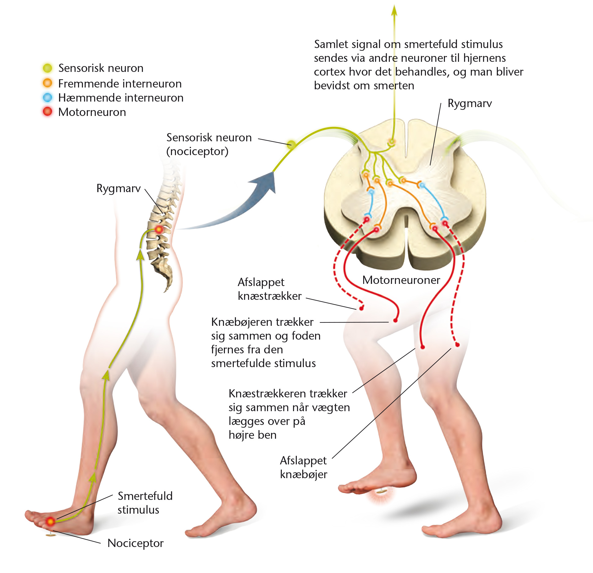
Figur 31
Afværgerefleks.
Illustration: Henning Dalhoff
ISBN 978-87-90363-84-0 · © Nucleus Forlag ApS.

Figur 32
Knæstrækkerrefleksen.
Illustration: Henning Dalhoff
ISBN 978-87-90363-84-0 · © Nucleus Forlag ApS.

Figur 33
Hjernens blodkar er omgivet af gliaceller.
Illustration: Henning Dalhoff
ISBN 978-87-90363-84-0 · © Nucleus Forlag ApS.

Figur 34
Membranpotentialet opstår gradvist ud fra en koncentrationsgradient.
Illustration: Henning Dalhoff
ISBN 978-87-90363-84-0 · © Nucleus Forlag ApS.

Figur 35
a. Cellemembran uden et membranpotentiale.
b. Cellemembran med membranpotentiale.
Illustration: Henning Dalhoff
ISBN 978-87-90363-84-0 · © Nucleus Forlag ApS.
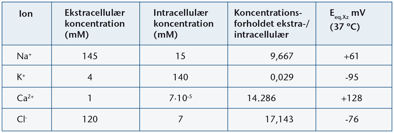
Figur 36
Typiske koncentrationer i intra- og ekstracellulærvæskerne.
Illustration: Birthe Møller Nielsen
ISBN 978-87-90363-84-0 · © Nucleus Forlag ApS.

Figur 38
Eksempler på forskellige transmitterstoffer.
Illustration: Elin Steffensen, Griffle
ISBN 978-87-90363-84-0 · © Nucleus Forlag ApS.

Figur 37
Synapse.
Illustration: Henning Dalhoff
ISBN 978-87-90363-84-0 · © Nucleus Forlag ApS.

Figur 42
Rumlig og tidsmæssig summation af postsynaptiske potentialer (PSP´er).
Illustration: Henning Dalhoff
ISBN 978-87-90363-84-0 · © Nucleus Forlag ApS.

Figur 39
Dannelse af det postsynaptiske potentiale (PSP).
Illustration: Henning Dalhoff
ISBN 978-87-90363-84-0 · © Nucleus Forlag ApS.

Figur 40
Receptorstyrede ionkanaler.
Illustration: Henning Dalhoff
ISBN 978-87-90363-84-0 · © Nucleus Forlag ApS.

Figur 41
Det exciterede postsynaptiske potentiale (EPSP).
Illustration: Henning Dalhoff
ISBN 978-87-90363-84-0 · © Nucleus Forlag ApS.

Figur 43
Den spændingsstyrede Na+-kanal i forskellige faser.
Illustration: Henning Dalhoff
ISBN 978-87-90363-84-0 · © Nucleus Forlag ApS.

Figur 45
Sammenhæng mellem aktionspotentiale, de spædingsstyrende ionkanaler og ionstrømninger-
Illustration: Henning Dalhoff
ISBN 978-87-90363-84-0 · © Nucleus Forlag ApS.

Figur 46
a. Etablering af længdegående ladningsstrømme.
b. Refraktærperiode.
Illustration: Elin Steffensen, Griffle
ISBN 978-87-90363-84-0 · © Nucleus Forlag ApS.

Figur 47
Saltatorisk eller springende impulsledning.
Illustration: Elin Steffensen, Griffle.
ISBN 978-87-90363-84-0 · © Nucleus Forlag ApS.

Figur 48
Tværsnit af hjernen.
Illustration: Henning Dalhoff
ISBN 978-87-90363-84-0 · © Nucleus Forlag ApS.

Figur 49
b. Kaffebær.
Illustration: Henning Dalhoff
ISBN 978-87-90363-84-0 · © Nucleus Forlag ApS.

Figur 50
Den kemiske formel for koffein.
ISBN 978-87-90363-84-0 · © Nucleus Forlag ApS.

Figur 51
Adenosins påvirkning af de postsynaptiske membraner.
Illustration: Henning Dalhoff
ISBN 978-87-90363-84-0 · © Nucleus Forlag ApS.

Figur 52
Koffein virker som antagonist til adenosin.
Illustration: Henning Dalhoff
ISBN 978-87-90363-84-0 · © Nucleus Forlag ApS.

Figur 53
Smagsløg med smagsceller.
Illustration: Henning Dalhoff
ISBN 978-87-90363-84-0 · © Nucleus Forlag ApS.

Figur 54
Registrering af surt og salt, sødt, bitter og umami.
Illustration: Henning Dalhoff
ISBN 978-87-90363-84-0 · © Nucleus Forlag ApS.

Figur 55
Smagspapiller med smagsløg.
Illustration: Henning Dalhoff
ISBN 978-87-90363-84-0 · © Nucleus Forlag ApS.

Figur 56
a. Næsen med lugtekolbe.
b. Olfaktorisk epithel.
Illustration: Henning Dalhoff
ISBN 978-87-90363-84-0 · © Nucleus Forlag ApS.

Figur 58
Transport af smerter.
Illustration: Henning Dalhoff
ISBN 978-87-90363-84-0 · © Nucleus Forlag ApS.

Figur 59
Smerteimpulsen kommer ind til rygraden.
Illustration: Henning Dalhoff
ISBN 978-87-90363-84-0 · © Nucleus Forlag ApS.

Figur 60
Smerteimpulsen stoppes af de endogene opioider.
Illustration: Henning Dalhoff
ISBN 978-87-90363-84-0 · © Nucleus Forlag ApS.

Figur 61
Oversigt over luftvejene.
Illustration: Henning Dalhoff
ISBN 978-87-90363-84-0 · © Nucleus Forlag ApS.

Figur 62
Cillier, også kaldet fimrehår, bevæger slimlag med partikler.
Illustration: Henning Dalhoff
ISBN 978-87-90363-84-0 · © Nucleus Forlag ApS.

Figur 63
De fineste bronkioler ender i alveolerne.
Illustration: Henning Dalhoff
ISBN 978-87-90363-84-0 · © Nucleus Forlag ApS.

Figur 64
Surfaktantmolekylerne forhindrer at alveolerne klapper sammen.
Illustration: Henning Dalhoff
ISBN 978-87-90363-84-0 · © Nucleus Forlag ApS.

Figur 65
De vigtigste muskler i brug under ind- og udånding.
Illustration: Henning Dalhoff
ISBN 978-87-90363-84-0 · © Nucleus Forlag ApS.

Figur 66
a. Person koblet til et vippespirometer.
b. Spirogram.
Illustration: Henning Dalhoff
ISBN 978-87-90363-84-0 · © Nucleus Forlag ApS.

Figur 67
Typiske værdier for lungernes forskellige voluminer.
Illustration: Birthe Møller Nielsen
ISBN 978-87-90363-84-0 · © Nucleus Forlag ApS.

Figur 68
Det døde rum.
Illustration: Henning Dalhoff
ISBN 978-87-90363-84-0 · © Nucleus Forlag ApS.

Figur 69
Diffusionsvejen fra alveolerne til kapillærerne er kort.
Illustration: Henning Dalhoff
ISBN 978-87-90363-84-0 · © Nucleus Forlag ApS.

Figur 70
Partialtrykket for O2 og CO2 i kredsløbets forskellige afsnit i hvile.
Illustration: Henning Dalhoff
ISBN 978-87-90363-84-0 · © Nucleus Forlag ApS.

Figur 71
Oxygenbindingskurver for hæmoglobin og myoglobin.
ISBN 978-87-90363-84-0 · © Nucleus Forlag ApS.

Figur 72
a. Hæmoglobin.
ISBN 978-87-90363-84-0 · © Nucleus Forlag ApS.

Figur 72
b. Hæmgruppe.
Illustration: Birthe Møller Nielsen
ISBN 978-87-90363-84-0 · © Nucleus Forlag ApS.

Figur 73
Ved samme pO2 opnår HbF en højere O2-mætning end HbA.
Illustration: Elin Steffensen, Griffle
ISBN 978-87-90363-84-0 · © Nucleus Forlag ApS.

Figur 74
Hæmoglobins evne til at binde O2 afhænger af pH.
Illustration: Erik Hjørne
ISBN 978-87-90363-84-0 · © Nucleus Forlag ApS.

Figur 75
Blodets afgivelse af O2 og CO2 i vævsvæske og -celler.
Illustration: Henning Dalhoff
ISBN 978-87-90363-84-0 · © Nucleus Forlag ApS.

Figur 76
Sanseceller i aortabue, halsarterier og den forlængde marv registrerer blodets pO2, pCO2 og pH.
Illustration: Henning Dalhoff
ISBN 978-87-90363-84-0 · © Nucleus Forlag ApS.

Figur 78
pO2 og pCO2 i blodet under et sikkert dyk.
Illustration: Elin Steffensen, Griffle
ISBN 978-87-90363-84-0 · © Nucleus Forlag ApS.

Figur 79
pO2 og pCO2 i blodet under et dyk efter hyperventilation.
Illustration: Elin Steffensen, Griffle
ISBN 978-87-90363-84-0 · © Nucleus Forlag ApS.

Figur 81
Nødvendigheden af at følge et dekompressionsskema.
Illustration: Elin Steffensen, Griffle
ISBN 978-87-90363-84-0 · © Nucleus Forlag ApS.

Figur 82
Oxygenlagre samt mængden af myoglobin i forskellige dykkende dyr.
Illustration: Birthe Møller Nielsen
ISBN 978-87-90363-84-0 · © Nucleus Forlag ApS.

Figur 83
Ændringer i en sæls kredsløb, når den den dykker og restituerer efter dykket.
Illustration: Elin Steffensen, Griffle
ISBN 978-87-90363-84-0 · © Nucleus Forlag ApS.

Figur 84
Koncentration af lactat i blodet efterendt dyk.
Illustration: Elin Steffensen, Griffle
ISBN 978-87-90363-84-0 · © Nucleus Forlag ApS.

Figur 87
Maksimal oxygenoptagelseshastighed hos bjergbestigere ved stigende højde.
Illustration: Elin Steffensen, Griffle
ISBN 978-87-90363-84-0 · © Nucleus Forlag ApS.

Figur 88
Blodkredsløb hos forskellige dyr.
a. Snegl. b. Haj. c. Frø
Illustration: Henning Dalhoff
ISBN 978-87-90363-84-0 · © Nucleus Forlag ApS.

Figur 89
Blodkredsløbet.
Illustration: Henning Dalhoff
ISBN 978-87-90363-84-0 · © Nucleus Forlag ApS.

Figur 90
Portåresystemet.
Illustration: Henning Dalhoff
ISBN 978-87-90363-84-0 · © Nucleus Forlag ApS.

Figur 91
Venepumpen.
Illustration: Henning Dalhoff
ISBN 978-87-90363-84-0 · © Nucleus Forlag ApS.

Figur 92
Hjertets opbygning.
Illustration: Henning Dalhoff
ISBN 978-87-90363-84-0 · © Nucleus Forlag ApS.

Figur 93
Blodforsyningen til hjertet.
Illustration: Henning Dalhoff
ISBN 978-87-90363-84-0 · © Nucleus Forlag ApS.

Figur 94
Hjertets kontraktioner.
Illustration: Henning Dalhoff
ISBN 978-87-90363-84-0 · © Nucleus Forlag ApS.

Figur 95
Elektrokardiogram, EKG.
ISBN 978-87-90363-84-0 · © Nucleus Forlag ApS.

Figur 96
Typiske eksempler på indre diameter og vægtykkelse i blodkarrene.
ISBN 978-87-90363-84-0 · © Nucleus Forlag ApS.

Figur 97
Blodets bestanddele.
Illustration: Henning Dalhoff
ISBN 978-87-90363-84-0 · © Nucleus Forlag ApS.

Figur 98
Stofudveksling og vandbevægelse gennem kapillærvæggen.
Illustration: Henning Dalhoff
ISBN 978-87-90363-84-0 · © Nucleus Forlag ApS.

Figur 99
Blodtrykkets fald i karssytemet jo længere man kommer fra hjertet.
Illustration: Elin Steffensen, Griffle
ISBN 978-87-90363-84-0 · © Nucleus Forlag ApS.

Figur 100
Hjertet under kontraktion.
ISBN 978-87-90363-84-0 · © Nucleus Forlag ApS.

Figur 101
Regulering af blodtrykket.
Illustration: Henning Dalhoff
ISBN 978-87-90363-84-0 · © Nucleus Forlag ApS.

Figur 102
Regulering af hjertets minutvolumen.
Illustration: Henning Dalhoff
ISBN 978-87-90363-84-0 · © Nucleus Forlag ApS.

Figur 103
Effekt af træning.
Illustration: Henning Dalhoff
ISBN 978-87-90363-84-0 · © Nucleus Forlag ApS.

Figur 106
Et gennemskåret hjerte.
Illustration: Henning Dalhoff
ISBN 978-87-90363-84-0 · © Nucleus Forlag ApS.

Figur 107
Eksempler på musklernes udseende.
Illustration: Henning Dalhoff
ISBN 978-87-90363-84-0 · © Nucleus Forlag ApS.

Figur 108
Oversigt over nogle af kroppens store skeletmuskler.
Illustration: Henning Dalhoff
ISBN 978-87-90363-84-0 · © Nucleus Forlag ApS.

Figur 109
Tværsnit gennem en muskel.
Illustration: Henning Dalhoff
ISBN 978-87-90363-84-0 · © Nucleus Forlag ApS.

Figur 110
Et lille udsnit af en gennemskåret muskelfiber.
Illustration: Henning Dalhoff
ISBN 978-87-90363-84-0 · © Nucleus Forlag ApS.

Figur 111
Stamceller deler sig under fosterudviklingen og udvikles til myoblaster som igen deler sig.
Illustration: Henning Dalhoff
ISBN 978-87-90363-84-0 · © Nucleus Forlag ApS.

Figur 112
a. Skitse af muskelfiber-tværsnit.
Illustration: Henning Dalhoff
ISBN 978-87-90363-84-0 · © Nucleus Forlag ApS.

Figur 113
b. Skematisk oversigt.
c. Opbygningen af myosin- og aktinfilamenter.
Illustration: Henning Dalhoff
ISBN 978-87-90363-84-0 · © Nucleus Forlag ApS.

Figur 114
Sarkomerstruktur med tværsnit tre forskellige steder.
Illustration: Henning Dalhoff
ISBN 978-87-90363-84-0 · © Nucleus Forlag ApS.

Figur 115
a. Aktinfilament - uden Ca2+ til stede.
b. Når Ca2+ bindes til troponin.
Illustration: Henning Dalhoff
ISBN 978-87-90363-84-0 · © Nucleus Forlag ApS.

Figur 116
Filamentforskydning.
Illustration: Henning Dalhoff
ISBN 978-87-90363-84-0 · © Nucleus Forlag ApS.

Figur 117
a. Et motorisk neuron danner synapser med en række muskelfibre.
b. Det motoriske neurons endeknopper danner synapser med den enkelte muskelfiber.
c. Forstørrelse af synapsen.
Illustration: Henning Dalhoff
ISBN 978-87-90363-84-0 · © Nucleus Forlag ApS.

Figur 118
Aktionspotentialet i muskelfiberen.
Illustration: Henning Dalhoff
ISBN 978-87-90363-84-0 · © Nucleus Forlag ApS.

Figur 119
Tværbrocyklus.
Illustration: Henning Dalhoff
ISBN 978-87-90363-84-0 · © Nucleus Forlag ApS.

Figur 120
Filamentforskydningen under en muskelkontraktion.
Illustration: Henning Dalhoff
ISBN 978-87-90363-84-0 · © Nucleus Forlag ApS.

Figur 121
Aktionspotentiale og muskelkraft.
Illustration: Elin Steffensen, Griffle
ISBN 978-87-90363-84-0 · © Nucleus Forlag ApS.

Figur 122
Kraftudvikling ved forskellige nerveimpulsfrekvenser.
a. Ved lav frekvens.
b-d. Ved højere frekvenser.
Illustration: Elin Steffensen, Griffle.
ISBN 978-87-90363-84-0 · © Nucleus Forlag ApS.

Figur 123
Antagonistiske muskler.
Illustration: Henning Dalhoff
ISBN 978-87-90363-84-0 · © Nucleus Forlag ApS.

Figur 124
Statisk og dynamisk arbejde.
Illustration: Henning Dalhoff
ISBN 978-87-90363-84-0 · © Nucleus Forlag ApS.

Figur 125
Muskelfiberens energiproduktion.
Illustration: Henning Dalhoff
ISBN 978-87-90363-84-0 · © Nucleus Forlag ApS.

Figur 126
Skematisk fremstilling af omtrentig procentfordeling af energibidrag.
ISBN 978-87-90363-84-0 · © Nucleus Forlag ApS.

Figur 128
Eksempler på bidrag til genopbygning af ATP under forskellige aktiviteter.
ISBN 978-87-90363-84-0 · © Nucleus Forlag ApS.

Figur 129
Oxygenforbrug og energileverance.
ISBN 978-87-90363-84-0 · © Nucleus Forlag ApS.

Figur 131
Kraftudvikling som funktion af tiden ved en enkeltkontraktion.
Illustration: Birthe Møller Nielsen
ISBN 978-87-90363-84-0 · © Nucleus Forlag ApS.

Figur 132
Kraftudvikling som funktion af tiden ved summerede kontraktioner.
Illustration: Erik Hjørne
ISBN 978-87-90363-84-0 · © Nucleus Forlag ApS.

Figur 133
Egenskaber for skeletmusklernes tre muskelfibertyper.
ISBN 978-87-90363-84-0 · © Nucleus Forlag ApS.

Figur 134
a. Kraftudvikling som funktion af kontraktionshastigheden.
b. Effekt som funktion af kontraktionshastigheden.
Illustration: Erik Hjørne
ISBN 978-87-90363-84-0 · © Nucleus Forlag ApS.

Figur 135
Fibertypesammensætning hos racer af hunde og heste i muskler involveret i løb.
ISBN 978-87-90363-84-0 · © Nucleus Forlag ApS.

Figur 136
Procentvis fordeling af type I- og type II-fibre i lårmuskel.
ISBN 978-87-90363-84-0 · © Nucleus Forlag ApS.

Figur 137
Ændringer i muskelfiberstørrelse.
ISBN 978-87-90363-84-0 · © Nucleus Forlag ApS.

Figur 138
a. Immunforsvaret efter hård og langvarig aktivitet.
b. Arbejdsintensitet og risiko for infektion.
Illustration: Erik Hjørne
ISBN 978-87-90363-84-0 · © Nucleus Forlag ApS.

Figur 139
Den arbejdende muskel udskiller IL-6.
Illustration: Henning Dalhoff
ISBN 978-87-90363-84-0 · © Nucleus Forlag ApS.

Figur 140
Børnedødelighed forårsaget af mæslinger.
Illustration: Elin Steffensen, Griffle
ISBN 978-87-90363-84-0 · © Nucleus Forlag ApS.

Figur 141
WHO´s faktaark om mæslinger.
Illustration: Birthe Møller Nielsen
ISBN 978-87-90363-84-0 · © Nucleus Forlag ApS.

Figur 142
Bakterie.
Illustration: Henning Dalhoff
ISBN 978-87-90363-84-0 · © Nucleus Forlag ApS.

Figur 143
Cellevæg hos grampositiv og gramnegativ bakterie.
Illustration: Henning Dalhoff
ISBN 978-87-90363-84-0 · © Nucleus Forlag ApS.

Figur 144
Virus uden og med kappe.
Illustration: Henning Dalhoff
ISBN 978-87-90363-84-0 · © Nucleus Forlag ApS.

Figur 146
Tårevæsken.
Illustration: Henning Dalhoff
ISBN 978-87-90363-84-0 · © Nucleus Forlag ApS.

Figur 147
Fimrehår bevæger slimlag med partikler.
Illustration: Henning Dalhoff
ISBN 978-87-90363-84-0 · © Nucleus Forlag ApS.

Figur 148
Immunforsvarets uspecifikke og specifikke del.
Illustration: Henning Dalhoff
ISBN 978-87-90363-84-0 · © Nucleus Forlag ApS.

Figur 149
Blodcelleproduktion.
Illustration: Henning Dalhoff
ISBN 978-87-90363-84-0 · © Nucleus Forlag ApS.

Figur 150
Granulocyt forlader blodbanen.
Illustration: Henning Dalhoff
ISBN 978-87-90363-84-0 · © Nucleus Forlag ApS.

Figur 151
Fagocytose af bakterier.
Illustration: Henning Dalhoff
ISBN 978-87-90363-84-0 · © Nucleus Forlag ApS.

Figur 152
Komplementsystemets vigtigste funktioner.
ISBN 978-87-90363-84-0 · © Nucleus Forlag ApS.

Figur 153
a. En virusinficeret celle.
b. En makrofag.
Illustration: Henning Dalhoff
ISBN 978-87-90363-84-0 · © Nucleus Forlag ApS.

Figur 154
Immunforsvaret ved en virusinfektion.
Illustration: Henning Dalhoff
ISBN 978-87-90363-84-0 · © Nucleus Forlag ApS.

Figur 155
En T-dræbercelle angriber en anden celle.
Illustration: Henning Dalhoff
ISBN 978-87-90363-84-0 · © Nucleus Forlag ApS.

Figur 156
Antistofklassers typer af lange kæder.
ISBN 978-87-90363-84-0 · © Nucleus Forlag ApS.

Figur 157
De fem antistofklasser.
Illustration: Henning Dalhoff
ISBN 978-87-90363-84-0 · © Nucleus Forlag ApS.

Figur 158
Sammenklumpning af antistoffer bundet til virus.
Illustration: Henning Dalhoff
ISBN 978-87-90363-84-0 · © Nucleus Forlag ApS.

Figur 159
Antistofrespons med primær og sekundær stimulering.
Illustration: Erik Hjørne
ISBN 978-87-90363-84-0 · © Nucleus Forlag ApS.

Figur 160
Antistofproduktion hos en allergiker.
Illustration: Henning Dalhoff
ISBN 978-87-90363-84-0 · © Nucleus Forlag ApS.

Figur 161
Allergen bundet til mastcelle udskiller histamin.
Illustration: Henning Dalhoff
ISBN 978-87-90363-84-0 · © Nucleus Forlag ApS.

Figur 164
Vækstkurve for sygdomsfremkaldende mikroorganismer.
Illustration: Elin Steffensen, Griffle
ISBN 978-87-90363-84-0 · © Nucleus Forlag ApS.

Figur 165
Influenzavirus.
Illustration: Henning Dalhoff
ISBN 978-87-90363-84-0 · © Nucleus Forlag ApS.

Figur 166
Infektionsforløb med influenzavirus.
Illustration: Henning Dalhoff
ISBN 978-87-90363-84-0 · © Nucleus Forlag ApS.

Figur 167
Fugleinfluenza.
Illustration: Henning Dalhoff
ISBN 978-87-90363-84-0 · © Nucleus Forlag ApS.

Figur 168
Koncentration af antistof i blodet.
Illustration: Elin Steffensen, Griffle
ISBN 978-87-90363-84-0 · © Nucleus Forlag ApS.

Figur 169
DNA-vaccination.
Illustration: Henning Dalhoff
ISBN 978-87-90363-84-0 · © Nucleus Forlag ApS.

Figur 173
ELISA til påvisning af henholdsvis antigener og antistoffer i patientprøver.
Illustration: Henning Dalhoff
ISBN 978-87-90363-84-0 · © Nucleus Forlag ApS.

Figur 174
Immunologiske test.
ISBN 978-87-90363-84-0 · © Nucleus Forlag ApS.

Figur 176
Parakrin signalering.
Illustration: Henning Dalhoff
ISBN 978-87-90363-84-0 · © Nucleus Forlag ApS.

Figur 177
Endokrin signalering.
Illustration: Henning Dalhoff
ISBN 978-87-90363-84-0 · © Nucleus Forlag ApS.

Figur 178
Målceller har receptorer der passer til det pågældende hormon.
Illustration: Henning Dalhoff
ISBN 978-87-90363-84-0 · © Nucleus Forlag ApS.

Figur 179
Rumlig opbygning af adrenalinreceptor skal passe til rumlig opbygning af adrenalinmolekyle.
Illustration: Henning Dalhoff
ISBN 978-87-90363-84-0 · © Nucleus Forlag ApS.

Figur 180
Menneskets hormonproducerende kirtler.
Illustration: Henning Dalhoff
ISBN 978-87-90363-84-0 · © Nucleus Forlag ApS.

Figur 181
Positiv feedback.
Illustration: Elin Steffensen, Griffle
ISBN 978-87-90363-84-0 · © Nucleus Forlag ApS.

Figur 182
Negativ feedback.
Illustration: Elin Steffensen, Griffle
ISBN 978-87-90363-84-0 · © Nucleus Forlag ApS.

Figur 183
Membranbundet hormonreceptor.
Illustration: Henning Dalhoff
ISBN 978-87-90363-84-0 · © Nucleus Forlag ApS.

Figur 184
Fedtopløselige hormoner bindes til intracellulære receptorer.
Illustration: Henning Dalhoff
ISBN 978-87-90363-84-0 · © Nucleus Forlag ApS.

Figur 185
Steroider.
Illustration: Birthe Møller Nielsen
ISBN 978-87-90363-84-0 · © Nucleus Forlag ApS.

Figur 186
Signaloverførsel gennrem en kaskadereaktion.
Illustration: Elin Steffensen, Griffle
ISBN 978-87-90363-84-0 · © Nucleus Forlag ApS.

Figur 187
Tyrosinkinasereceptor.
Illustration: Henning Dalhoff
ISBN 978-87-90363-84-0 · © Nucleus Forlag ApS.

Figur 188
G-protein-receptor.
Illustration: Henning Dalhoff
ISBN 978-87-90363-84-0 · © Nucleus Forlag ApS.

Figur 189
Steroidhormoner bindes til intracellulære receptorer.
Illustration: Henning Dalhoff
ISBN 978-87-90363-84-0 · © Nucleus Forlag ApS.

Figur 196
Bestøvning af æbleblomst og livscyklus.
Illustration: Henning Dalhoff
ISBN 978-87-90363-84-0 · © Nucleus Forlag ApS.

Figur 197
Kønsdifferentiering hos foster.
Illustration: Henning Dalhoff
ISBN 978-87-90363-84-0 · © Nucleus Forlag ApS.

Figur 198
Y-kromosom med SRY-gen.
ISBN 978-87-90363-84-0 · © Nucleus Forlag ApS.

Figur 199
Udvikling af kønskirtler hos fostre.
Illustration: Henning Dalhoff
ISBN 978-87-90363-84-0 · © Nucleus Forlag ApS.

Figur 200
Kvindens kønsorganer.
Illustration: Henning Dalhoff
ISBN 978-87-90363-84-0 · © Nucleus Forlag ApS.

Figur 201
Hormonel påvirkning af pubertetsudviklingen.
Illustration: Henning Dalhoff
ISBN 978-87-90363-84-0 · © Nucleus Forlag ApS.

Figur 202
Pubertetsændringer hos piger.
Illustration: Erik Hjørne
ISBN 978-87-90363-84-0 · © Nucleus Forlag ApS.

Figur 203
Menestruationscyklus.
Illustration: Henning Dalhoff
ISBN 978-87-90363-84-0 · © Nucleus Forlag ApS.

Figur 205
Meiose hos mænd og kvinder.
Illustration: Henning Dalhoff
ISBN 978-87-90363-84-0 · © Nucleus Forlag ApS.

Figur 206
a. Befrugtning i æggeleder.
Illustration: Henning Dalhoff
ISBN 978-87-90363-84-0 · © Nucleus Forlag ApS.

Figur 206
b. Æggets udvikling
Illustration: Henning Dalhoff
ISBN 978-87-90363-84-0 · © Nucleus Forlag ApS.

Figur 207
Hormonelle ændringer under graviditet.
Illustration: Birthe Møller Nielsen
ISBN 978-87-90363-84-0 · © Nucleus Forlag ApS.

Figur 208
Oversigt over hormoner under graviditet.
ISBN 978-87-90363-84-0 · © Nucleus Forlag ApS.

Figur 209
Moderkagens funktion.
ISBN 978-87-90363-84-0 · © Nucleus Forlag ApS.

Figur 210
Mandens kønsorganer.
a. Tværsnit set fra siden.
b. Tværsnit af penis.
Illustration: Henning Dalhoff
ISBN 978-87-90363-84-0 · © Nucleus Forlag ApS.

Figur 211
Snit gennem testikel.
Illustration: Henning Dalhoff
ISBN 978-87-90363-84-0 · © Nucleus Forlag ApS.

Figur 212
Tværsnit af sædkanal.
Illustration: Henning Dalhoff
ISBN 978-87-90363-84-0 · © Nucleus Forlag ApS.

Figur 213
Pubertetsændringer hos drenge.
Illustration: Erik Hjørne
ISBN 978-87-90363-84-0 · © Nucleus Forlag ApS.

Figur 214
Hormonregulering hos mænd.
Illustration: Henning Dalhoff
ISBN 978-87-90363-84-0 · © Nucleus Forlag ApS.

Figur 215
Østrogenlignende stoffer hæmmer sædcelleproduktionen.
Illustration: Henning Dalhoff
ISBN 978-87-90363-84-0 · © Nucleus Forlag ApS.

Figur 216
Den hormonelle regulering under stress.
Illustration: Henning Dalhoff
ISBN 978-87-90363-84-0 · © Nucleus Forlag ApS.

Figur 217
Virkning af adrenalin og noradrenalin.
Illustration: Elin Steffensen, Griffle.
ISBN 978-87-90363-84-0 · © Nucleus Forlag ApS.

Figur 218
Oversigt over forskellige hormoner.
Illustration: Birthe Møller Nielsen
ISBN 978-87-90363-84-0 · © Nucleus Forlag ApS.

Figur 219
a. Monosaccharider.
ISBN 978-87-90363-84-0 · © Nucleus Forlag ApS.

Figur 219
b. Disaccharider.
ISBN 978-87-90363-84-0 · © Nucleus Forlag ApS.

Figur 219
c. Polysaccharider.
ISBN 978-87-90363-84-0 · © Nucleus Forlag ApS.

Figur 220
a. Sammensætning af aminosyrer.
b. Tripeptid dannet ud fra tre aminosyrer.
ISBN 978-87-90363-84-0 · © Nucleus Forlag ApS.

Figur 221
a. Triglycerid, fedtsyre og glycerol.
Illustration: Erik Hjørne
ISBN 978-87-90363-84-0 · © Nucleus Forlag ApS.

Figur 221
b. Phospholipid.
ISBN 978-87-90363-84-0 · © Nucleus Forlag ApS.

Figur 221
c. Cholesterol.
Illustration: Birthe Møller Nielsen
ISBN 978-87-90363-84-0 · © Nucleus Forlag ApS.

Figur 222
Mættet, monoumættet og polyumættet fedtsyre.
Illustration: Birthe Møller Nielsen
ISBN 978-87-90363-84-0 · © Nucleus Forlag ApS.

Figur 225
Oversigt over fordøjelsessystemet.
Illustration: Henning Dalhoff
ISBN 978-87-90363-84-0 · © Nucleus Forlag ApS.

Figur 226
Fordøjelsessystemet.
Illustration: Henning Dalhoff
ISBN 978-87-90363-84-0 · © Nucleus Forlag ApS.

Figur 227
Spytkirtlernes placering.
Illustration: Henning Dalhoff
ISBN 978-87-90363-84-0 · © Nucleus Forlag ApS.

Figur 228
Peristaltisk bevægelse i spiserøret.
Illustration: Henning Dalhoff
ISBN 978-87-90363-84-0 · © Nucleus Forlag ApS.

Figur 229
Påvirkning får hjernen til at sende besked til mavesækken om dannelse af gastrin.
Illustration: Henning Dalhoff
ISBN 978-87-90363-84-0 · © Nucleus Forlag ApS.

Figur 230
Yderligere dannelse af mavesaft og gastrin.
Illustration: Henning Dalhoff
ISBN 978-87-90363-84-0 · © Nucleus Forlag ApS.

Figur 231
Negativ feedbackmekanisme.
Illustration: Henning Dalhoff
ISBN 978-87-90363-84-0 · © Nucleus Forlag ApS.

Figur 232
Pepsin spalter proteiners peptidbindinger.
ISBN 978-87-90363-84-0 · © Nucleus Forlag ApS.

Figur 233
Hormonel regulering af bugskpytkirtlens udskillelse af HCO3-.
Illustration: Henning Dalhoff
ISBN 978-87-90363-84-0 · © Nucleus Forlag ApS.

Figur 234
Oversigt over fordøjelsessystemets hormoner.
Illustration: Birthe Møller Nielsen
ISBN 978-87-90363-84-0 · © Nucleus Forlag ApS.

Figur 235
Hormonel regulering af bugspytkirtlens udskillelse af enzymer.
Illustration: Henning Dalhoff
ISBN 978-87-90363-84-0 · © Nucleus Forlag ApS.

Figur 236
Galdeblærens og bugspytkirtlens fælles udmunding i tolvfingertarmen.
Illustration: Henning Dalhoff
ISBN 978-87-90363-84-0 · © Nucleus Forlag ApS.

Figur 237
Udskillelse af de proteinspaltende enzymer fra bugspytkirtlen.
Illustration: Henning Dalhoff
ISBN 978-87-90363-84-0 · © Nucleus Forlag ApS.

Figur 238
Tyndtarmens opbygning.
Illustration: Henning Dalhoff
ISBN 978-87-90363-84-0 · © Nucleus Forlag ApS.

Figur 239
Fordøjelse af stivelse i tyndtarmen.
Illustration: Henning Dalhoff
ISBN 978-87-90363-84-0 · © Nucleus Forlag ApS.

Figur 240
Spaltning af disaccharider og dextriner i tyndtarmens slimhinde.
Illustration: Henning Dalhoff
ISBN 978-87-90363-84-0 · © Nucleus Forlag ApS.

Figur 241
Optagelse af monosacchariderne.
Illustration: Henning Dalhoff
ISBN 978-87-90363-84-0 · © Nucleus Forlag ApS.

Figur 242
Aminosyrer og små peptider binder sig til samme transportproteiner.
Illustration: Henning Dalhoff
ISBN 978-87-90363-84-0 · © Nucleus Forlag ApS.

Figur 243
Nedbrydning og optagelse af triglycerid.
Illustration: Henning Dalhoff
ISBN 978-87-90363-84-0 · © Nucleus Forlag ApS.

Figur 244
Spaltning af triglycerid.
ISBN 978-87-90363-84-0 · © Nucleus Forlag ApS.

Figur 245
Optagelsesmekanismer for forskellige mineraler i tarmsystemet.
Illustration: Henning Dalhoff
ISBN 978-87-90363-84-0 · © Nucleus Forlag ApS.

Figur 246
Fordøjelseskanalens væske- og ionbalance.
Illustration: Henning Dalhoff
ISBN 978-87-90363-84-0 · © Nucleus Forlag ApS.

Figur 247
Koleratoksinet forårsager diarré.
Illustration: Henning Dalhoff
ISBN 978-87-90363-84-0 · © Nucleus Forlag ApS.

Figur 250
Hjerne-tarm-aksen.
Illustration: Henning Dalhoff
ISBN 978-87-90363-84-0 · © Nucleus Forlag ApS.

Figur 253
Energiomsætningens komponenter.
ISBN 978-87-90363-84-0 · © Nucleus Forlag ApS.

Figur 254
Beregning af basalstofskiftet.
Illustration: Birthe Møller Nielsen
ISBN 978-87-90363-84-0 · © Nucleus Forlag ApS.

Figur 255
Gennemsnitlig energiforbrug.
ISBN 978-87-90363-84-0 · © Nucleus Forlag ApS.

Figur 256
a. Bugspytkirtel.
b. Binyre.
c. Hypofyse.
Illustration: Henning Dalhoff
ISBN 978-87-90363-84-0 · © Nucleus Forlag ApS.

Figur 257
Insulinreceptor.
Illustration: Henning Dalhoff
ISBN 978-87-90363-84-0 · © Nucleus Forlag ApS.

Figur 258
Transport af glucose ind i celle.
Illustration: Henning Dalhoff
ISBN 978-87-90363-84-0 · © Nucleus Forlag ApS.

Figur 259
Transportører af glucose og andre monosaccharider.
Illustration: Birthe Møller Nielsen
ISBN 978-87-90363-84-0 · © Nucleus Forlag ApS.

Figur 260
Carbohydratomsætningen efter et måltid.
Illustration: Henning Dalhoff
ISBN 978-87-90363-84-0 · © Nucleus Forlag ApS.

Figur 262
Proteinomsætningen efter et måltid.
Illustration: Henning Dalhoff
ISBN 978-87-90363-84-0 · © Nucleus Forlag ApS.
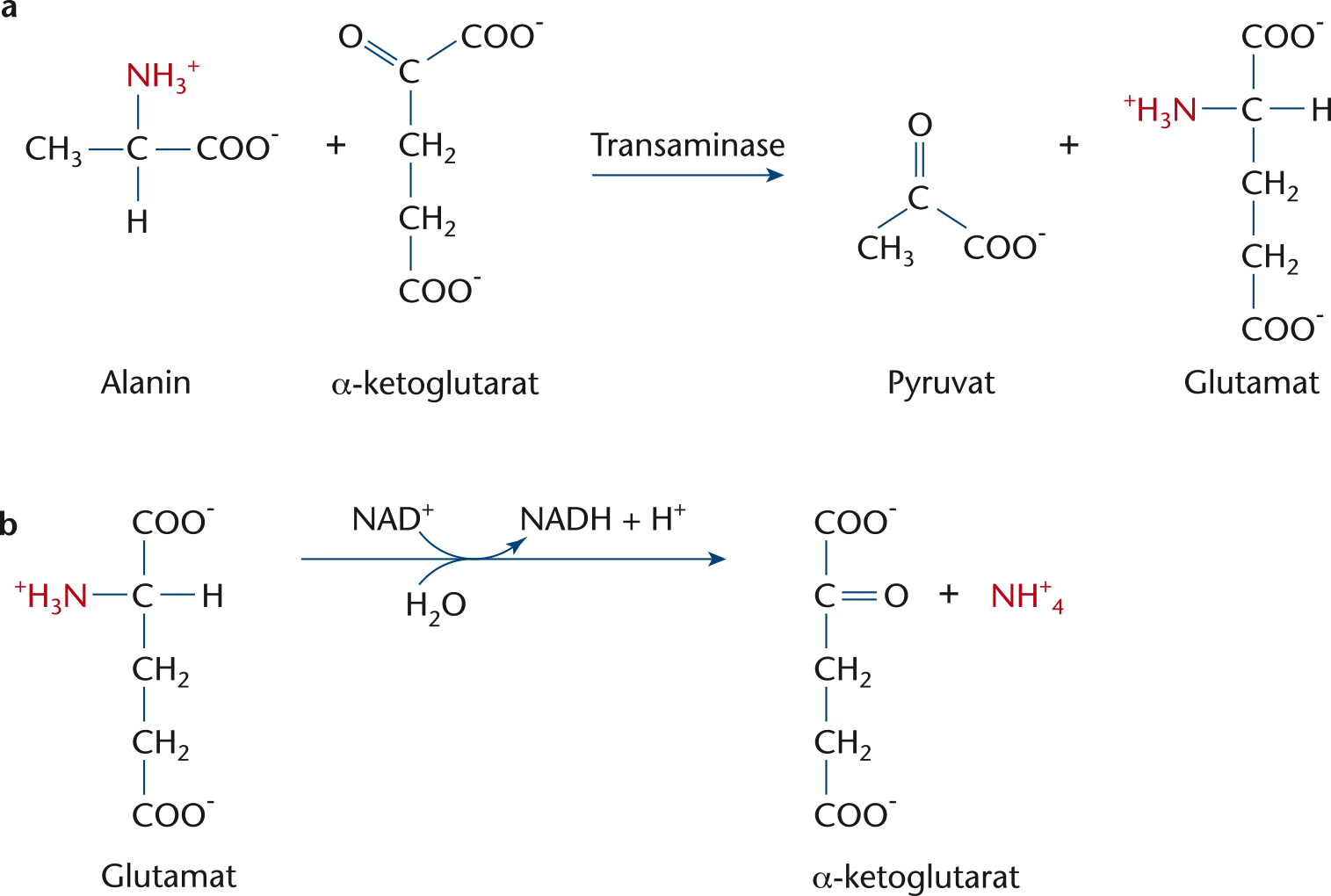
Figur 263
a. Transaminering.
b. Deaminering.
ISBN 978-87-90363-84-0 · © Nucleus Forlag ApS.

Figur 264
a. Triglycerid.
ISBN 978-87-90363-84-0 · © Nucleus Forlag ApS.

Figur 264
b. Phospholipid.
ISBN 978-87-90363-84-0 · © Nucleus Forlag ApS.

Figur 264
c. Cholesterol.
Illustration: Birthe Møller Nielsen
ISBN 978-87-90363-84-0 · © Nucleus Forlag ApS.

Figur 265
Lipoproteinomsætningen.
Illustration: Henning Dalhoff
ISBN 978-87-90363-84-0 · © Nucleus Forlag ApS.

Figur 266
Lipidomsætningen efter et måltid.
Illustration: Henning Dalhoff
ISBN 978-87-90363-84-0 · © Nucleus Forlag ApS.

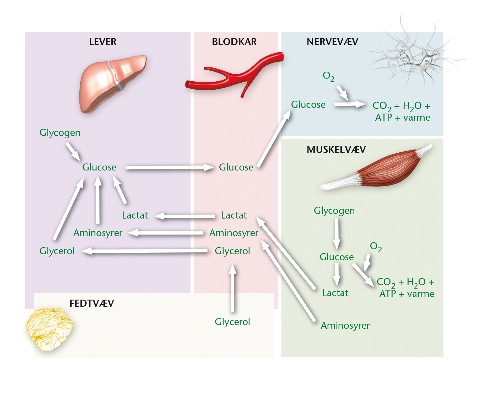
Figur 267
Processer i lever-, muskel- og fedtvæv.
Illustration: Henning Dalhoff
ISBN 978-87-90363-84-0 · © Nucleus Forlag ApS.
Figur 268
Carbohydratomsætning mellem måltider.
Illustration: Henning Dalhoff
ISBN 978-87-90363-84-0 · © Nucleus Forlag ApS.

Figur 269
Proteinomsætning mellem måltider.
Illustration: Henning Dalhoff
ISBN 978-87-90363-84-0 · © Nucleus Forlag ApS.

Figur 270
Lipidomsætning mellem måltider.
Illustration: Henning Dalhoff
ISBN 978-87-90363-84-0 · © Nucleus Forlag ApS.

Figur 271
Næringsstofomsætning mellem måltider.
Illustration: Henning Dalhoff
ISBN 978-87-90363-84-0 · © Nucleus Forlag ApS.

Figur 272
Processer der stimuleres af glucagon, adrenalin, væksthormon og cortisol.
Illustration: Henning Dalhoff
ISBN 978-87-90363-84-0 · © Nucleus Forlag ApS.

Figur 273
Sult- og mæthedssignaler.
Illustration: Henning Dalhoff
ISBN 978-87-90363-84-0 · © Nucleus Forlag ApS.

Figur 274
Sammenligning af diabetes type 1 og type 2.
Illustration: Birthe Møller Nielsen
ISBN 978-87-90363-84-0 · © Nucleus Forlag ApS.

Figur 275
Alkohols omsætning i kroppen.
ISBN 978-87-90363-84-0 · © Nucleus Forlag ApS.

Figur 276
16-24-åriges alkoholforbrug.
Illustration: Birthe Møller Nielsen
ISBN 978-87-90363-84-0 · © Nucleus Forlag ApS.

Figur 278
Omsætning af paracetamol.
ISBN 978-87-90363-84-0 · © Nucleus Forlag ApS.

Figur 279
Udvikling af overvægt.
Illustration: Elin Steffensen, Griffle
ISBN 978-87-90363-84-0 · © Nucleus Forlag ApS.

Figur 280
Sammehæng mellem BMI og dødelighed forårsaget af hjertekarsygdomme.
ISBN 978-87-90363-84-0 · © Nucleus Forlag ApS.

Figur 281
THR, Talje-Hofte-Ratio.
Illustration: Henning Dalhoff
ISBN 978-87-90363-84-0 · © Nucleus Forlag ApS.

Figur 283
Sammenhæng mellem fysisk aktivitet, BMI og udvikling af diabetes type 2.
Illustration: Elin Steffensen, Griffle
ISBN 978-87-90363-84-0 · © Nucleus Forlag ApS.

Figur 284
Metabolisk syndrom og stigende fysisk aktivitet.
Illustration: Elin Steffensen, Griffle
ISBN 978-87-90363-84-0 · © Nucleus Forlag ApS.